The structure of the human respiratory system presentation. Presentation for the lesson "Breathing, its meaning. The structure and function of the respiratory system." presentation for a lesson in biology (grade 8) on the topic. III. Learning new material
Respiratory structure
Biology teacher MBOU SOSH number 8
Krasny Sulin
Truscheleva Svetlana Semyonovna

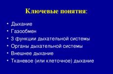
Key concepts :
- Breath
- Gas exchange
- 3 functions of the respiratory system
- Respiratory system organs
- External respiration
- Tissue (or cellular) respiration

What do I know on this topic
New, unknown

Breathing is the exchange of gases between the body and the environment.
The breathing process consists
from 4 stages:
- the exchange of gases between the air and the lungs;
- the exchange of gases between the lungs and blood;
- transport of gases by blood;
- gas exchange in tissues.
Respiratory system
performs only the first part
gas exchange. Rest
is performed by the organ system
blood circulation. Between
respiratory and circulatory
systems, there is a deep
interconnection.

Gas exchange
Gas exchange in the lungs (external respiration)
Gas exchange in tissues (cellular respiration)

- The human respiratory organs, according to their functional characteristics, can be divided into two groups: air, or respiratory and gas exchange organs. : nasal cavity → nasopharynx → larynx → trachea → bronchi. Gas exchange organs: lungs.

- Providing gas exchange
- Participate in heat regulation (when breathing, water evaporates from the surface of the lungs, which leads to cooling of the blood and the whole body)
- Voice formation (the lungs create air currents that vibrate the vocal cords of the larynx).

- Air purification
- Air humidification
- Air disinfection
- Warming the air
- Perception of smells (organ of smell)

- Functions:
- breathing protection of the lower respiratory tract voice production
- breath
- lower respiratory tract protection
- voice formation
- Located at level IV-VI of the cervical vertebrae
- The entrance to the larynx is protected by a special semi-movable cartilage - the epiglottis

The thyroid cartilage in men protrudes somewhat forward, forming an Adam's apple. The vocal cords are located in the narrow part of the larynx.

Trachea and bronchi - organs of the lower respiratory tract
Trachea
Structure: a wide tube 9-11 cm long, consisting of 16-20 cartilaginous half-rings on the soft side facing the esophagus. The inner wall of the trachea is covered with ciliated epithelium.
Functions: free passage of air into the lungs, removal of pollen particles from the lungs into the pharynx.
Bronchi
Structure: branching tubes of a smaller diameter. They consist of cartilaginous rings that protect them from falling off during inhalation.
Functions: Air supply to the alveoli of the lungs.

Lungs
Each lung is covered with a membrane - the pulmonary pleura. The thoracic cavity is also lined by a membrane - the parietal pleura. Between the parietal and pulmonary pleura, there is a narrow gap - the pleural cavity filled with the thinnest layer of fluid, which facilitates the sliding of the pulmonary wall during inhalation and exhalation.

Human lungs consist of the smallest pulmonary vesicles - alveoli
The alveoli are braided by a network of blood vessels - capillaries. The alveoli are formed by the epithelium, which secretes a special fluid, the thinnest film lining the alveoli (surfactant). Its functions: reduces surface tension and prevents the alveoli from closing; kills germs that have entered the lungs. In the alveoli, gas is exchanged between the blood and the surrounding air by diffusion.

Gas exchange in tissues
The oxygen content in the tissue fluid is lower than in the arterial blood; therefore, oxygen from the capillaries enters the tissue fluid. From it, it diffuses into the cells, where it immediately enters into energy exchange reactions (oxidizes organic compounds and releases energy), therefore, there is almost no free oxygen in the cells.
In the reactions of energy metabolism, carbon dioxide is formed. Its concentration in the cells becomes higher than in the tissue fluid, and the gas diffuses into it, and then to the capillaries. In them, one part of the carbon dioxide molecules dissolves in the blood plasma, while the other enters the erythrocytes.

- P. 158-161 textbook
- Creative:
- - compose a crossword puzzle on the topic
- - make a presentation "Sound Shaping"

Lesson objectives:
- Educational:
- to study the structural features of the respiratory organs in connection with their functions;
- to reveal the essence of the breathing process, its importance in metabolism;
- find out the mechanisms of voice formation;
- Developing:
- continue the formation of the basics of hygiene (breathing hygiene rules);
- develop research skills through the setting of educational experiments;
- Educational:
- to educate a respectful attitude towards your body, towards your health, towards the health of others;
- draw an analogy: breath is life; human lungs are the lungs of our planet (flora).
A healthy planet is a healthy person!
DURING THE CLASSES
I. Organizational moment
II. Updating basic knowledge
It is possible to show a fragment of a video film on the topic.
- What is breathing?
- Does the structure of the organ affect the function it performs?
We will try to find answers to all these and many other questions in today's lesson.
III. Learning new material
Appendix. Slide number 7.
Respiratory system
consists of airways(cavities and tubes connected in series) and respiratory part.
TO airways includes the nasal cavity and nasopharynx (upper respiratory tract), larynx, trachea and bronchi.
Respiratory part- this is the lungs and the connective tissue sheath - the pleura.
Appendix. Slide number 8.
Respiratory system
- Here is a table that we will try to fill in while studying new material. Redraw it, please. (It is better to print and distribute the table in advance, so as not to waste precious lesson time on this)
Appendix. Slide number 9.
Upper respiratory tract
During normal breathing, air must pass through the external nostrils into the nasal cavity, which is divided into two halves by the bone-cartilaginous septum. In each half there are sinuous nasal passages that increase the surface of the nasal cavity. Their walls are lined with a mucous membrane containing numerous cells of the ciliated (ciliated) epithelium.
In an adult, the mucous membrane secretes 0.5 liters of mucus per day.
Its function is to humidify the inhaled air, trap dust particles and microorganisms that settle on the walls of the cavity. The mucus contains substances that kill microbes or prevent them from reproducing (the enzyme lysozyme and leukocytes). Numerous blood vessels branch out under the mucous membrane, so even minor injuries to the nose are accompanied by profuse bleeding. These choroid plexuses warm the inhaled air to body temperature. The nasal cavity is connected to the cavities in the bones of the skull: maxillary, frontal and wedge-shaped. They serve not only to warm the incoming air, but also serve as resonators for voice formation. The nasal cavities are lined with sensitive cells that provide a protective function: the sneezing reflex. The nasal cavity opens into the nasopharynx with the internal nostrils - the choans, and from there - into the larynx.
Appendix. Slide number 10. Hygiene of nasal breathing
- It is recommended to breathe through the nose, because when breathing through the mouth, cold air enters the lungs, which is the cause of colds.
- A sick person who does not follow the rules of hygiene becomes a source of infection.
(After explaining the structure and functions of a separate organ, you can check the correctness of filling in the table, or you can single out this as an independent work as a consolidation of the material or as homework)
Appendix. Slide number 11.
Observations
"Check the air passage through the nasal passages"
We close one nasal passage, and bring a light piece of cotton wool to the other. The jet of air will throw it away when you exhale, and press it against the nasal opening when you inhale. This technique can be shown on the subject.
Conclusion: During normal breathing, air must pass through the external nostrils into the nasal cavity.
Appendix. Slide number 12.
Larynx
The larynx is like a funnel, the walls of which are formed by cartilage.
The larynx cavity is lined with a mucous membrane and equipped with receptors - a reflex cough.
The entrance to the larynx during swallowing is closed by the supraglottic cartilage.
The largest cartilage is thyroid, which protects the larynx in front.
The vocal cords are stretched between the cartilages, and the glottis is located between the ligaments.
Thus, the function of the larynx is to conduct air into the trachea, participate in voice formation and prevent the penetration of harmful substances into the respiratory tract.
Appendix. Slide number 13.
Observation
1. To prove that when swallowing, the thyroid cartilage rises up.
Feel for the thyroid cartilage, swallow. Make sure that the cartilage goes up and then returns to its original place.
Conclusion: with this movement, the epiglottis closes the entrance to the trachea and along it, like a bridge, the saliva or food lump moves into the esophagus.
2. Find out why breathing stops during swallowing.
Make another swallowing motion and make sure that this fact is true.
Conclusion: the tongue closes the entrance to the nasal cavity, the epiglottis blocks the entrance to the trachea. As a result, air cannot enter the lungs at the time of swallowing.
Appendix. Slide number 14.
Sound production
The person is silent - the glottis is triangular and large enough.
The sound appears when the glottis is incompletely closed, air passes through it, which vibrates the vocal cords.
The shorter the vocal cords, the higher the sound. The final formation of sound occurs in the cavities of the pharynx, nasopharynx, mouth and nose (remember the sinuses?) And depends on the position of the lips, lower jaw and tongue.
Appendix. Slide number 15.
Phonogram of the word MAMA, which clearly shows that consonants cause a stronger vibration of the vocal cords than vowels.
Appendix. Slide number 16. Hygiene of the vocal apparatus
Screaming damages the vocal cords, which can cause inflammation, hoarseness, or loss of voice. When whispering, the ligaments relax and do not close completely. Frequent inflammation of the respiratory tract, smoking and alcohol have a negative effect on the voice-forming apparatus.
Appendix. Slide number 17
Trachea and bronchi
The larynx, a 10–12 cm tube, passes directly into the trachea, which is in front of the esophagus. Its anterior wall is formed by cartilaginous half rings, so the lumen of the trachea is always open.
The back wall is soft and adjacent to the esophagus.
At the bottom, the trachea is divided into 2 bronchi. Both the trachea and the bronchi are lined by the mucous membrane, which contains ciliated epithelium with glandular cells. Here the air is saturated with water vapor and purified.
Appendix. Slide number 18. Breathing hygiene
- Swallowing large pieces of food can choke and block the trachea.
- In inflammatory conditions, a cough occurs, which helps to remove mucus from the airways.
Appendix. Slide number 19
Lungs
The lungs are a large paired cone-shaped organ. Outside covered with pulmonary pleura; the chest cavity is covered with parietal pleura, between them there is a pleural cavity that does not contain air. It is filled with liquid, which reduces breathing friction. 100 liters of air pass through the lungs in 1 minute. What is the structure of the lung?
Appendix. Slide number 20.
Internal structure of the lung
The bronchi, entering the lungs, continue to branch, forming bronchioles, at the ends of which there are clusters of thin-walled pulmonary vesicles - alveoli. The walls of the alveoli and capillaries are single-layered, which facilitates gas exchange. The cells of the epithelium of the alveoli secrete biologically active substances that form a surfactant, which prevents the adhesion of the alveoli and neutralizes microorganisms that have entered the lungs.
The spent surfactant is digested by phagocytes or excreted as sputum.
Appendix. Slide number 21.Breathing hygiene
With pulmonary diseases, surfactant may not be released, then the alveoli are closed and do not participate in gas exchange. Smoking interferes with the physiological properties of the surfactant.
Appendix. Slide number 22It is interesting
- 300-350 million alveoli with a total area of 100 square meters
- Pulmonary capillary length - 7-8 microns
- Blood passes through the capillaries of the alveoli in 0.8 s, but the hemoglobin has time to be saturated with oxygen
Appendix. Slide number 23
Observation
Find out how full breathing differs from shallow breathing.
Do you know how to breathe correctly? It turns out that this is very important, especially in winter and during the transitional winter-spring period, during a flu epidemic. According to experts, improper breathing significantly increases the likelihood of respiratory pathogens entering the body, which increases the risk of getting the flu or colds.
Many people breathe too often (and the norm is 16 breaths per minute in a calm state) and shallow, from time to time holding their breath in and out. This type of breathing is called shallow breathing. As a result, the lungs do not have time to properly ventilate - fresh air enters only the outer sections, while most of the lung volume remains, as it were, unclaimed, that is, the air in it is not renewed. And viruses and bacteria just need it.
Full breath is a combination of lower, middle and upper breathing. A person who practices full breathing all the time will have a wide chest - and any narrow-chested person can develop their chest to a normal size.
Let's check if you are breathing correctly. To do this, put a watch with a second hand in front of you, sit comfortably, relax, straighten your shoulders. Count how many breaths you take in a minute. Follow the rhythm of breathing: the ratio of inhalation and exhalation, the arrangement of pauses in this cycle. Determine exactly how you breathe: actively relaxing the abdomen - abdominal breathing, raising and lowering the chest - chest type, combining both - mixed breathing.
If you are taking less than 14 breaths per minute, great. Well trained and hardy people usually breathe this way. You can be rightfully proud of yourself. Taking in the air deeply, you allow your lungs to expand, ventilate them perfectly, that is, you make your respiratory system almost invulnerable to infectious agents.
A good result is considered to be from 14 to 18 breaths per minute. This is how most practically healthy people breathe, who can get flu or ARVI no more than 2 times a season.
More than 18 breaths per minute is already a serious cause for concern. With shallow and frequent breathing, only half of the inhaled air enters the lungs. This is clearly not enough for the constant renewal of the pulmonary atmosphere.
Appendix. Slide number 24 and 25. test yourself(material fixing)
It is necessary to connect the organ and the function it performs with arrows. This table can be printed to check that each student has completed it correctly.
Appendix. Slide number 26. test yourself(material fixing)
- Let's return to the questions that were posed at the beginning of the lesson and try to answer them.
- What is breathing?
- Why do they say: breathing is life?
- Does the structure of the organ affect the function it performs? Etc.
(According to the proposed drawings, each teacher will be able to formulate their own questions depending on the preparation of the class and on the amount of time remaining, etc.)
Appendix. Slide number 27.Homework
Creative laboratory:
1. In what cases is nasal breathing difficult? What are the consequences of this violation? Offer a code of practice for breathing hygiene.
2. Develop recommendations and a set of exercises to correct breathing.


Basic terms and definitions: Breathing Breathing is a set of processes that ensure the supply of oxygen, its use in the oxidation of organic substances and the removal of carbon dioxide and some other substances. Respiratory Organs Respiratory organs are specialized organs for gas exchange between the body and the environment


The biological significance of respiration The biological significance of respiration: 1. Providing the body with oxygen. 2. Removal of carbon dioxide. 3. Oxidation of organic compounds BZHU with the release of energy necessary for a person to live. 4. Removal of end products of metabolism (water vapor, ammonia, hydrogen sulfide, etc.)

Respiratory system Respiratory part Airways of sequentially connected cavities and tubes: 1) nasal cavity, 2) nasopharynx, 3) larynx, 4) trachea 5) bronchi. the place where gas exchange occurs: 1) lungs 2) pleura - (connective tissue membrane)


Mln. alveoli with a total area of 100 sq. m 2. The length of the pulmonary capillary is 7-8 microns 3. Blood passes through the capillaries of the alveoli in 0.8 s, but the hemoglobin has time to be saturated with oxygen It's interesting:

Organ 1. Nasal cavity 2. Larynx 3. Trachea and bronchi 4. Lungs 5. Pulmonary and parietal pleura Function a) contains a fluid that reduces friction b) air humidification, dust retention c) provides free air passage d) formation of sounds, reflex cough e) gas exchange through the alveolar-capillary membrane Check yourself

Authority Function to be performed a) b) c) d) e) Check yourself

To use the preview of presentations, create yourself a Google account (account) and log into it: https://accounts.google.com
Slide captions:
With which organ system are the respiratory organs connected to perform the function of gas exchange?
Distribution of functions between the respiratory and circulatory systems Respiratory organs ensure the constancy of O 2 and CO 2 in the lungs by gas exchange between the air of the pulmonary alveoli and atmospheric air. The circulatory organs carry out gas exchange between the lungs and the blood, transfer O 2 to tissues and CO 2 from tissues to the lungs, and provide tissue gas exchange. The intensity of external respiration depends on the needs of the body, i.e. on the intensity of tissue gas exchange.
Respiration value 1. Providing the body with oxygen (O 2) 2. Oxidation (decomposition) of organic compounds with the release of energy 3. Formation and removal of excess carbon dioxide (CO 2) from the body 4. Removal of some end products of metabolism: water vapor (H 2 O), ammonia (NH 3), hydrogen sulfide (H 2 S), etc.
Breathing 1. External - gases diffuse through the respiratory surface of the lungs into the blood 2. Internal - gases diffuse between the circulating blood and the breathing cell 3. Tissue and cellular - occurs during the oxidation of nutrients to release energy: O 2 is consumed and CO2 is released
Respiratory system organs 1. Nasal cavity 2. Nasopharynx 3. Larynx 5. Bronchi 4. Trachea Lungs Airways 1 2 3 4 5 5 L L
Nasal cavity Sinus of the sphenoid bone Sinus of the frontal bone Nasopharynx Oropharynx Superior nasal concha Epiglottis Inferior turbinate Hard palate Soft palate
Functions of the nasal cavity 1. Warming (cooling) the inhaled air. 2. Humidification of the inhaled air. 3. Retaining and removing dust. 4. Destruction of bacteria. 5. Reflex sneezing. 7. Smell.
Larynx Epiglottis Thyroid cartilage Laryngeal cavity
Functions of the larynx 1. Formation of sounds and speech. 2. Reflex cough when the receptors are irritated from dust. 3. The epiglottis, when swallowing, closes the entrance to the larynx.
Sound production Air during exhalation Passes through the glottis Causes vibrations of the vocal cords Sound is produced
sound production Pitch length Soprano Bass The shorter the vocal cords, the higher their sound. The vibrational frequency of the ligaments is from 80 to 10,000 Hz.
Trachea and bronchi Trachea Bronchi
Functions of the trachea and bronchi Provide free passage of air
Lungs Right lung: 1,2,3 lobes. Left lung: 1.2 lobes. 1 2 3 1 2
Lung lobule diagram
Functions of the lungs 1. Gas exchange through the alveolo - capillary membrane. Epithelial cells secrete a surfactant substance, which prevents alveoli from sticking together and neutralizes microorganisms that have entered the lungs.
Pleura Each lung is covered with two sheets of connective tissue membrane: the pulmonary pleura is adjacent to the lungs, the parietal pleura - to the chest cavity. Between the pleural layers there is a pleural cavity filled with pleural fluid.
Answers 1. - F 2. - D 3. - A 4. - C 5. - D 6. - F 7. - B 8. - Z
Diffusion of gases in tissues O 2 O 2 O 2 O 2 O 2 CO 2 CO 2 CO 2 CO 2 capillary Tissue fluid Tissue fluid of the cell
Application: Larynx Larynx